Why 3D CBCT Imaging Is Essential for Safe Wisdom Teeth Removal in Kelowna
Why 3D CBCT Imaging Is Essential for Safe Wisdom Teeth Removal in Kelowna
Why 3D CBCT Imaging Is Essential for Safe Wisdom Teeth Removal in Kelowna

1. Introduction: The Evolution of Oral Surgery Diagnostics
Extracting third molars (wisdom teeth) is one of the most common surgical procedures in Canada. However, because wisdom teeth develop deep within the posterior mandible and maxilla, their proximity to vital anatomical structures demands precision diagnosis before any incision is made.
Historically, dentists relied exclusively on two-dimensional (2D) panoramic radiographs. While traditional 2D X-rays provide a helpful high-level overview, they flatten complex, three-dimensional facial structures into a flat image. This limitation introduces spatial distortion, structural overlap, and anatomical uncertainty.
Today, state-of-the-art 3D CBCT imaging for wisdom teeth removal in Kelowna has transformed modern oral surgery. Cone Beam Computed Tomography (CBCT) provides sub-millimeter 3D visual reconstruction of bone volume, tooth position, root curvature, and neurovascular pathways. At Kelowna Oral & Facial Surgery, integrating CBCT technology ensures every extraction is mapped with high anatomical accuracy, significantly reducing patient risks and accelerating post-operative recovery.
2. The Technological Gap: 2D Panoramic X-Rays vs. 3D CBCT Scanning
To understand why advanced imaging is essential for complex oral surgery, patients must understand how 2D radiographs differ from true 3D volumetric scans.
Diagnostic Feature | Traditional 2D Panoramic X-Ray | Advanced 3D CBCT Scan |
|---|---|---|
Dimensionality | Flat 2D representation | Full 3D volumetric visualization |
Anatomical Overlap | High (structures shadow one another) | Zero (layer-by-layer cross-sectional slicing) |
Nerve Localization | Estimated proximity only | Exact sub-millimeter 3D nerve mapping |
Root Curvature Mapping | Distorted angles often missed | Complete multi-planar root morphology |
Bone Density Assessment | Qualitative estimate | Quantitative bone height & quality measurement |
Radiation Dose | Low | Ultra-low (significantly lower than medical CTs) |
The Danger of 2D Structural Overlap
In a standard 2D panoramic radiograph, the roots of an impacted lower wisdom tooth often appear to overlap directly with the Inferior Alveolar Nerve (IAN) canal. However, a 2D image cannot reveal depth. It cannot answer critical surgical questions:
Is the tooth root located buccally (toward the cheek) or lingually (toward the tongue) relative to the nerve?
Is the root simply resting beside the nerve canal, or is it grooved, notched, or completely encircling the nerve tissue?
Attempting an extraction without answering these questions increases the risk of accidental nerve compression, stretching, or transection. 3D CBCT imaging for wisdom teeth removal in Kelowna eliminates this guesswork by offering multi-planar cross-sections (axial, coronal, and sagittal views), enabling oral surgeons to evaluate spatial relationships down to the fraction of a millimeter.
3. Protecting Vital Structures: Preventing Inferior Alveolar & Lingual Nerve Damage
The primary technical objective during lower wisdom tooth extraction is preserving the Inferior Alveolar Nerve (IAN) and the Lingual Nerve (LN). The IAN provides sensory innervation to the lower lip, chin, teeth, and gums on each side of the face. Damage to this nerve during surgery can lead to paresthesia (numbness), dysesthesia (painful burning sensations), or permanent sensory loss.
CBCT Diagnostic & Neuro-Mapping Workflow
Step 1: High-Resolution 3D Scan — Volumetric data acquisition of the jaw and surrounding facial structures.
Step 2: Neuro-Vascular Trace — Multi-planar slicing of the mandibular canal to pinpoint nerve pathways.
Step 3: Surgical Path Design — Custom tooth sectioning angles and safe removal vectors mapped around vital structures.
Step 4: Precision Execution — Minimally invasive extraction with targeted Inferior Alveolar Nerve preservation.
High-Risk Anatomical Scenarios Identified by CBCT
Nerve Canal Superimposition: When roots wrap around or contact the IAN canal, 3D imaging allows the surgeon to section the tooth crown and gently lift root fragments away from the nerve trajectory.
Sinus Perforation Prevention: Upper wisdom teeth often rest immediately against the floor of the Maxillary Sinus. A 3D scan provides exact measurements of the sinus membrane boundary, preventing accidental sinus communication or root displacement into the sinus cavity.
Ankylosis & Complex Root Curves: Hooked (dilacerated) roots or bone-fused (ankylosed) teeth are identified beforehand, allowing surgeons to design custom sectioning plans rather than applying excessive mechanical force.
By utilizing high-resolution 3D scanning, our surgical team transitions wisdom tooth removal from a traditional procedure to a highly predictable, targeted microsurgical operation.
4. The Surgical Advantage: Custom 3D Anatomical Modeling & Virtual Planning
At Kelowna Oral & Facial Surgery, patient care begins long before entering the operating suite. Utilizing modern 3D CBCT imaging data, specialized software generates a high-definition 3D virtual model of the patient’s facial skeleton.
Pre-Operative Virtual Plan Execution
Precision Tooth Sectioning: Rather than pulling an impacted tooth intact—which requires larger incisions and greater bone removal—3D modeling allows the oral surgeon to calculate precise sectioning angles. The tooth is systematically divided into micro-segments and removed with minimal force.
Bone Preservation: By assessing bone density and thickness surrounding the impacted tooth, surgeons cut only the absolute minimum amount of bone necessary, preserving structural mandibular integrity.
Reduced Surgical Time: Precise pre-operative planning streamlines the surgical process, reducing overall procedure time. Shorter operating times directly translate to reduced tissue exposure, lower infection risk, and reduced post-operative inflammation.
5. Technology Standards at Kelowna Oral & Facial Surgery
Providing safe wisdom teeth removal in Kelowna requires continuous investment in medical technology and surgical training. The imaging suite at Kelowna Oral & Facial Surgery utilizes ultra-low-dose CBCT technology (including the Vatech Green CT system) optimized specifically for the maxillofacial region.
Kelowna Oral & Facial Surgery Diagnostic Suite Capabilities
Ultra-Low-Dose CBCT Imaging:
Sub-millimeter diagnostic resolution
Extra-oral scanning comfort with no uncomfortable intra-oral films
Minimal radiation exposure adhering strictly to the ALARA (As Low As Reasonably Achievable) principle
Full 3D Neuro-Mapping:
Precise Inferior Alveolar and Lingual Nerve localization
Maxillary sinus boundary mapping
Quantitative bone quality and density metrics
Minimizing Radiation Exposure
Patients often express concern regarding radiation exposure during dental imaging. Modern CBCT technology operates on targeted, pulse-exposure protocols. A typical 3D CBCT scan of the jaws yields a radiation dose comparable to a few days of natural background radiation, and significantly less than a traditional medical-grade head CT scan. This makes 3D CBCT scanning an exceptionally safe diagnostic standard for adolescents, teenagers, and adult patients alike.
6. Clinical Authority: Led by Board-Certified Specialists Dr. Scott Martyna & Dr. Gordon Wong
Diagnostic technology is only as effective as the specialist interpreting the data. At Kelowna Oral & Facial Surgery, your care is directed by board-certified oral and maxillofacial surgeons Dr. Scott Martyna and Dr. Gordon Wong.
Surgical Leadership Team
Dr. Scott Martyna, DMD, MHA, FRCDC, FACS — Certified Specialist in Oral & Maxillofacial Surgery
Board-certified specialist in both Canada and the United States
Advanced surgical training and reconstructive maxillofacial procedures
Experienced in complex wisdom tooth extractions, digital treatment planning, and bone grafting techniques
Dr. Gordon Wong, DMD, FRCDC, Dip ABOMS — Certified Specialist in Oral & Maxillofacial Surgery
Board-certified specialist in both Canada and the United States
Extensive experience in 3D computer modeling, surgical simulation, and virtual surgical planning
Specialist in IV sedation, general anesthesia, and patient safety protocols
With decades of combined hospital and private-practice surgical experience—including hospital privileges at Kelowna General Hospital—Dr. Martyna and Dr. Wong specialize in evaluating complex CBCT scans, managing severe tooth impactions, and performing precise, minimally invasive extractions under IV sedation or general anesthesia. Their surgical expertise ensures that every patient in Kelowna receives hospital-grade safety standards within a modern, comfortable outpatient clinic environment.
7. Step-by-Step Surgical Timeline: What Patients Expect
From initial consultation to complete healing, our streamlined clinical protocol ensures safety, comfort, and peace of mind.
Overview of the 5-Step Surgical Process
Step 1: Initial Consultation & Ultra-Low-Dose 3D CBCT Scan
Step 2: Virtual Surgical Planning & Anesthesia Protocol Design
Step 3: Precision Surgical Removal (Under IV Sedation or General Anesthesia)
Step 4: Targeted Bone Grafting & Tissue Preservation (If Needed)
Step 5: Post-Operative Recovery Monitoring & Guided Healing
Stage 1: Consultation & High-Resolution Imaging
Upon arrival at our Kelowna clinic, patients undergo a rapid, non-invasive 3D CBCT scan. Dr. Martyna or Dr. Wong reviews the 3D anatomical model with the patient, pointing out tooth positioning, root orientation, and nerve pathways.
Stage 2: Anesthesia Selection & Surgical Preparation
Our facility offers comprehensive sedation options tailored to individual anxiety levels and surgical complexity, including local anesthesia, moderate IV sedation, and deep general anesthesia.
Stage 3: Targeted, Minimally Invasive Extraction
Using the 3D plan as a blueprint, the surgeon accesses the wisdom tooth through tiny micro-incisions. The tooth is sectioned smoothly along pre-calculated planes, protecting the surrounding bone and nerve tissue.
Stage 4: Rapid Post-Operative Recovery
Because bone and soft tissue disruption is kept to a minimum, patients experience reduced post-operative swelling, minimal pain, and a faster return to daily activities. Detailed home-care instructions and follow-up support are provided to ensure a smooth recovery.
8. Frequently Asked Questions (FAQs)
Why do I need a 3D CBCT scan if I already had a regular dental X-ray?
Regular dental X-rays produce flat 2D images that flatten complex facial structures. A 2D X-ray cannot show the exact depth, thickness, or 3D spatial relationship between your wisdom tooth roots and your nerve pathways. A 3D CBCT scan provides sub-millimeter cross-sectional views, allowing your surgeon to plan a safer extraction and avoid nerve injury.
Is the radiation from a 3D CBCT scan dangerous?
No. Modern CBCT units used at Kelowna Oral & Facial Surgery utilize focused, ultra-low-dose technology designed around the ALARA safety principle. The radiation exposure from a localized CBCT scan is a fraction of a traditional hospital CT scan and is comparable to a few days of normal background radiation from the environment.
Will insurance cover 3D CBCT imaging in Kelowna?
Many private dental insurance and medical plans cover diagnostic 3D imaging when deemed medically necessary for surgical procedures, such as impacted wisdom teeth near major nerve trunks. Our administrative team assists patients by completing and submitting the necessary insurance claim paperwork on your behalf to help maximize your reimbursement.
Does 3D imaging make wisdom tooth removal less painful?
Yes, indirectly. By using 3D imaging to plan precise tooth sectioning and micro-incisions, your oral surgeon minimizes bone removal and soft tissue trauma. Less physical trauma during surgery directly correlates with reduced post-operative pain, less swelling, and faster wound healing.
1. Introduction: The Evolution of Oral Surgery Diagnostics
Extracting third molars (wisdom teeth) is one of the most common surgical procedures in Canada. However, because wisdom teeth develop deep within the posterior mandible and maxilla, their proximity to vital anatomical structures demands precision diagnosis before any incision is made.
Historically, dentists relied exclusively on two-dimensional (2D) panoramic radiographs. While traditional 2D X-rays provide a helpful high-level overview, they flatten complex, three-dimensional facial structures into a flat image. This limitation introduces spatial distortion, structural overlap, and anatomical uncertainty.
Today, state-of-the-art 3D CBCT imaging for wisdom teeth removal in Kelowna has transformed modern oral surgery. Cone Beam Computed Tomography (CBCT) provides sub-millimeter 3D visual reconstruction of bone volume, tooth position, root curvature, and neurovascular pathways. At Kelowna Oral & Facial Surgery, integrating CBCT technology ensures every extraction is mapped with high anatomical accuracy, significantly reducing patient risks and accelerating post-operative recovery.
2. The Technological Gap: 2D Panoramic X-Rays vs. 3D CBCT Scanning
To understand why advanced imaging is essential for complex oral surgery, patients must understand how 2D radiographs differ from true 3D volumetric scans.
Diagnostic Feature | Traditional 2D Panoramic X-Ray | Advanced 3D CBCT Scan |
|---|---|---|
Dimensionality | Flat 2D representation | Full 3D volumetric visualization |
Anatomical Overlap | High (structures shadow one another) | Zero (layer-by-layer cross-sectional slicing) |
Nerve Localization | Estimated proximity only | Exact sub-millimeter 3D nerve mapping |
Root Curvature Mapping | Distorted angles often missed | Complete multi-planar root morphology |
Bone Density Assessment | Qualitative estimate | Quantitative bone height & quality measurement |
Radiation Dose | Low | Ultra-low (significantly lower than medical CTs) |
The Danger of 2D Structural Overlap
In a standard 2D panoramic radiograph, the roots of an impacted lower wisdom tooth often appear to overlap directly with the Inferior Alveolar Nerve (IAN) canal. However, a 2D image cannot reveal depth. It cannot answer critical surgical questions:
Is the tooth root located buccally (toward the cheek) or lingually (toward the tongue) relative to the nerve?
Is the root simply resting beside the nerve canal, or is it grooved, notched, or completely encircling the nerve tissue?
Attempting an extraction without answering these questions increases the risk of accidental nerve compression, stretching, or transection. 3D CBCT imaging for wisdom teeth removal in Kelowna eliminates this guesswork by offering multi-planar cross-sections (axial, coronal, and sagittal views), enabling oral surgeons to evaluate spatial relationships down to the fraction of a millimeter.
3. Protecting Vital Structures: Preventing Inferior Alveolar & Lingual Nerve Damage
The primary technical objective during lower wisdom tooth extraction is preserving the Inferior Alveolar Nerve (IAN) and the Lingual Nerve (LN). The IAN provides sensory innervation to the lower lip, chin, teeth, and gums on each side of the face. Damage to this nerve during surgery can lead to paresthesia (numbness), dysesthesia (painful burning sensations), or permanent sensory loss.
CBCT Diagnostic & Neuro-Mapping Workflow
Step 1: High-Resolution 3D Scan — Volumetric data acquisition of the jaw and surrounding facial structures.
Step 2: Neuro-Vascular Trace — Multi-planar slicing of the mandibular canal to pinpoint nerve pathways.
Step 3: Surgical Path Design — Custom tooth sectioning angles and safe removal vectors mapped around vital structures.
Step 4: Precision Execution — Minimally invasive extraction with targeted Inferior Alveolar Nerve preservation.
High-Risk Anatomical Scenarios Identified by CBCT
Nerve Canal Superimposition: When roots wrap around or contact the IAN canal, 3D imaging allows the surgeon to section the tooth crown and gently lift root fragments away from the nerve trajectory.
Sinus Perforation Prevention: Upper wisdom teeth often rest immediately against the floor of the Maxillary Sinus. A 3D scan provides exact measurements of the sinus membrane boundary, preventing accidental sinus communication or root displacement into the sinus cavity.
Ankylosis & Complex Root Curves: Hooked (dilacerated) roots or bone-fused (ankylosed) teeth are identified beforehand, allowing surgeons to design custom sectioning plans rather than applying excessive mechanical force.
By utilizing high-resolution 3D scanning, our surgical team transitions wisdom tooth removal from a traditional procedure to a highly predictable, targeted microsurgical operation.
4. The Surgical Advantage: Custom 3D Anatomical Modeling & Virtual Planning
At Kelowna Oral & Facial Surgery, patient care begins long before entering the operating suite. Utilizing modern 3D CBCT imaging data, specialized software generates a high-definition 3D virtual model of the patient’s facial skeleton.
Pre-Operative Virtual Plan Execution
Precision Tooth Sectioning: Rather than pulling an impacted tooth intact—which requires larger incisions and greater bone removal—3D modeling allows the oral surgeon to calculate precise sectioning angles. The tooth is systematically divided into micro-segments and removed with minimal force.
Bone Preservation: By assessing bone density and thickness surrounding the impacted tooth, surgeons cut only the absolute minimum amount of bone necessary, preserving structural mandibular integrity.
Reduced Surgical Time: Precise pre-operative planning streamlines the surgical process, reducing overall procedure time. Shorter operating times directly translate to reduced tissue exposure, lower infection risk, and reduced post-operative inflammation.
5. Technology Standards at Kelowna Oral & Facial Surgery
Providing safe wisdom teeth removal in Kelowna requires continuous investment in medical technology and surgical training. The imaging suite at Kelowna Oral & Facial Surgery utilizes ultra-low-dose CBCT technology (including the Vatech Green CT system) optimized specifically for the maxillofacial region.
Kelowna Oral & Facial Surgery Diagnostic Suite Capabilities
Ultra-Low-Dose CBCT Imaging:
Sub-millimeter diagnostic resolution
Extra-oral scanning comfort with no uncomfortable intra-oral films
Minimal radiation exposure adhering strictly to the ALARA (As Low As Reasonably Achievable) principle
Full 3D Neuro-Mapping:
Precise Inferior Alveolar and Lingual Nerve localization
Maxillary sinus boundary mapping
Quantitative bone quality and density metrics
Minimizing Radiation Exposure
Patients often express concern regarding radiation exposure during dental imaging. Modern CBCT technology operates on targeted, pulse-exposure protocols. A typical 3D CBCT scan of the jaws yields a radiation dose comparable to a few days of natural background radiation, and significantly less than a traditional medical-grade head CT scan. This makes 3D CBCT scanning an exceptionally safe diagnostic standard for adolescents, teenagers, and adult patients alike.
6. Clinical Authority: Led by Board-Certified Specialists Dr. Scott Martyna & Dr. Gordon Wong
Diagnostic technology is only as effective as the specialist interpreting the data. At Kelowna Oral & Facial Surgery, your care is directed by board-certified oral and maxillofacial surgeons Dr. Scott Martyna and Dr. Gordon Wong.
Surgical Leadership Team
Dr. Scott Martyna, DMD, MHA, FRCDC, FACS — Certified Specialist in Oral & Maxillofacial Surgery
Board-certified specialist in both Canada and the United States
Advanced surgical training and reconstructive maxillofacial procedures
Experienced in complex wisdom tooth extractions, digital treatment planning, and bone grafting techniques
Dr. Gordon Wong, DMD, FRCDC, Dip ABOMS — Certified Specialist in Oral & Maxillofacial Surgery
Board-certified specialist in both Canada and the United States
Extensive experience in 3D computer modeling, surgical simulation, and virtual surgical planning
Specialist in IV sedation, general anesthesia, and patient safety protocols
With decades of combined hospital and private-practice surgical experience—including hospital privileges at Kelowna General Hospital—Dr. Martyna and Dr. Wong specialize in evaluating complex CBCT scans, managing severe tooth impactions, and performing precise, minimally invasive extractions under IV sedation or general anesthesia. Their surgical expertise ensures that every patient in Kelowna receives hospital-grade safety standards within a modern, comfortable outpatient clinic environment.
7. Step-by-Step Surgical Timeline: What Patients Expect
From initial consultation to complete healing, our streamlined clinical protocol ensures safety, comfort, and peace of mind.
Overview of the 5-Step Surgical Process
Step 1: Initial Consultation & Ultra-Low-Dose 3D CBCT Scan
Step 2: Virtual Surgical Planning & Anesthesia Protocol Design
Step 3: Precision Surgical Removal (Under IV Sedation or General Anesthesia)
Step 4: Targeted Bone Grafting & Tissue Preservation (If Needed)
Step 5: Post-Operative Recovery Monitoring & Guided Healing
Stage 1: Consultation & High-Resolution Imaging
Upon arrival at our Kelowna clinic, patients undergo a rapid, non-invasive 3D CBCT scan. Dr. Martyna or Dr. Wong reviews the 3D anatomical model with the patient, pointing out tooth positioning, root orientation, and nerve pathways.
Stage 2: Anesthesia Selection & Surgical Preparation
Our facility offers comprehensive sedation options tailored to individual anxiety levels and surgical complexity, including local anesthesia, moderate IV sedation, and deep general anesthesia.
Stage 3: Targeted, Minimally Invasive Extraction
Using the 3D plan as a blueprint, the surgeon accesses the wisdom tooth through tiny micro-incisions. The tooth is sectioned smoothly along pre-calculated planes, protecting the surrounding bone and nerve tissue.
Stage 4: Rapid Post-Operative Recovery
Because bone and soft tissue disruption is kept to a minimum, patients experience reduced post-operative swelling, minimal pain, and a faster return to daily activities. Detailed home-care instructions and follow-up support are provided to ensure a smooth recovery.
8. Frequently Asked Questions (FAQs)
Why do I need a 3D CBCT scan if I already had a regular dental X-ray?
Regular dental X-rays produce flat 2D images that flatten complex facial structures. A 2D X-ray cannot show the exact depth, thickness, or 3D spatial relationship between your wisdom tooth roots and your nerve pathways. A 3D CBCT scan provides sub-millimeter cross-sectional views, allowing your surgeon to plan a safer extraction and avoid nerve injury.
Is the radiation from a 3D CBCT scan dangerous?
No. Modern CBCT units used at Kelowna Oral & Facial Surgery utilize focused, ultra-low-dose technology designed around the ALARA safety principle. The radiation exposure from a localized CBCT scan is a fraction of a traditional hospital CT scan and is comparable to a few days of normal background radiation from the environment.
Will insurance cover 3D CBCT imaging in Kelowna?
Many private dental insurance and medical plans cover diagnostic 3D imaging when deemed medically necessary for surgical procedures, such as impacted wisdom teeth near major nerve trunks. Our administrative team assists patients by completing and submitting the necessary insurance claim paperwork on your behalf to help maximize your reimbursement.
Does 3D imaging make wisdom tooth removal less painful?
Yes, indirectly. By using 3D imaging to plan precise tooth sectioning and micro-incisions, your oral surgeon minimizes bone removal and soft tissue trauma. Less physical trauma during surgery directly correlates with reduced post-operative pain, less swelling, and faster wound healing.
Other blogs

Dental Implants vs. Bone Grafting: Rebuilding Your Jaw Foundation in Kelowna

Anesthesia & Sedation Options at Kelowna Oral & Facial Surgery: What to Expect
Why 3D CBCT Imaging Is Essential for Safe Wisdom Teeth Removal in Kelowna

Fractured Teeth and the Importance of Dental Crowns

Dental X-Rays -> Types and why are they Important?

Periodontal Disease: Why Routfine Care Is Essential?

Transforming Your Smile and Oral Health
Contact Information
Leave Us a Message
Parking Instructions
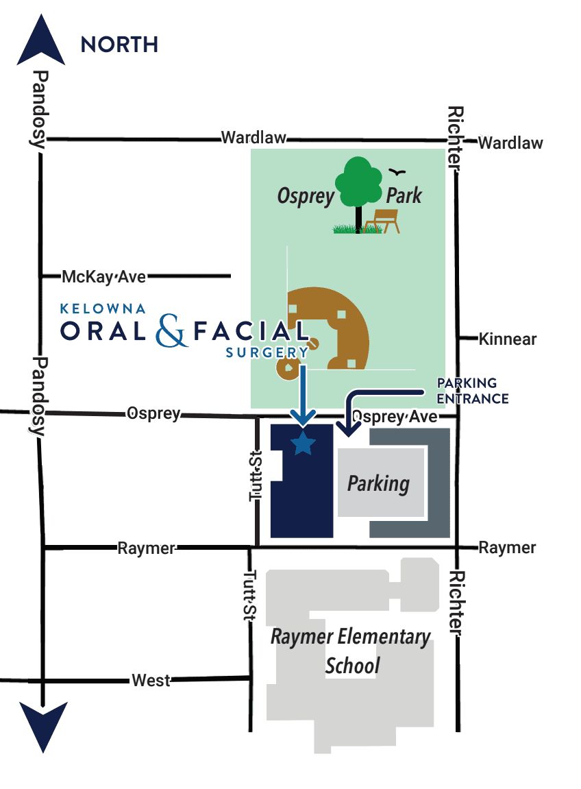
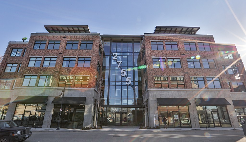
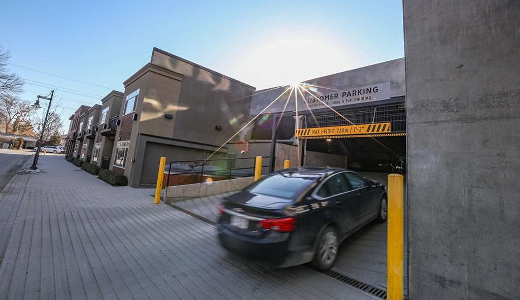
The parkade is linked to our building and the entrance to the garage is on Osprey Avenue. Please bring your license plate number with you so we can register your vehicle upon arrival.
Contact Information
Leave Us a Message
Parking Instructions
The parkade is linked to our building and the entrance to the garage is on Osprey Avenue. Please bring your license plate number with you so we can register your vehicle upon arrival.
Contact Information
Leave Us a Message
Parking Instructions
The parkade is linked to our building and the entrance to the garage is on Osprey Avenue. Please bring your license plate number with you so we can register your vehicle upon arrival.

Copyright © 2026 Dr. Scott T. Martyna, DMD, MHA, FRCDC, FACS, & Dr. Gordon Wong, DMD, FRCDC, Dip ABOMS Certified Specialists in Oral & Maxillofacial Surgery. All rights reserved.
Kelowna Oral & Facial Surgery is locally owned and operated by Dr. Martyna and Dr. Wong Dental Inc.
Copyright © 2026 Dr. Scott T. Martyna, DMD, MHA, FRCDC, FACS, & Dr. Gordon Wong, DMD, FRCDC, Dip ABOMS Certified Specialists in Oral & Maxillofacial Surgery. All rights reserved.
Kelowna Oral & Facial Surgery is locally owned and operated by Dr. Martyna and Dr. Wong Dental Inc.
Copyright © 2026 Dr. Scott T. Martyna, DMD, MHA, FRCDC, FACS, & Dr. Gordon Wong, DMD, FRCDC, Dip ABOMS Certified Specialists in Oral & Maxillofacial Surgery. All rights reserved.
Kelowna Oral & Facial Surgery is locally owned and operated by Dr. Martyna and Dr. Wong Dental Inc.